

Diabetic Retinopathy Detection
- Availibility: In Stock
1. Hardware Requirements:
-
Camera/Imaging Device:
- Fundus Camera: High-resolution retinal camera capable of capturing detailed images of the retina.
- Optical Coherence Tomography (OCT) (optional): For cross-sectional images of the retina.
-
Computing Hardware:
- Microcontroller/Processor: High-performance CPU or GPU, such as Intel Core i7 or NVIDIA GPU, for processing and training deep learning models.
- Storage: Sufficient storage for images and models, e.g., SSD with 1TB capacity.
- Memory: At least 16GB RAM for processing large datasets.
-
Connectivity:
- Internet: For cloud storage, model training, and data upload.
- Network Interface: Ethernet or Wi-Fi for connectivity.
2. Software Requirements:
-
Operating System:
- Linux or Windows (preferably Linux for server deployment).
-
Programming Languages:
- Python: For data processing, model training, and inference.
- JavaScript/HTML/CSS: For web-based user interface (if applicable).
-
Libraries and Frameworks:
- TensorFlow or PyTorch: For deep learning and model training.
- OpenCV: For image preprocessing and computer vision tasks.
- scikit-learn: For additional machine learning algorithms.
- Keras: High-level API for building and training neural networks (optional if using TensorFlow).
-
Development Tools:
- Jupyter Notebook or Google Colab: For experimentation and model development.
- IDE: Integrated Development Environment like PyCharm or Visual Studio Code.
3. Data Specifications:
-
Dataset:
- Images: High-resolution retinal images, typically fundus images.
- Annotations: Labels for different stages of diabetic retinopathy (e.g., No DR, Mild DR, Moderate DR, Severe DR, Proliferative DR).
- Format: Common formats include JPEG, PNG, TIFF.
-
Preprocessing:
- Normalization: Rescale pixel values to [0, 1] or [-1, 1].
- Augmentation: Techniques like rotation, scaling, and flipping to increase dataset diversity.
4. Model Specifications:
-
Architecture:
- Convolutional Neural Network (CNN): A CNN model with layers such as Conv2D, MaxPooling2D, Flatten, and Dense.
- Pre-trained Models (optional): Models like ResNet, Inception, or VGG can be used as a starting point.
-
Training:
- Loss Function: Categorical Cross-Entropy for multi-class classification.
- Optimizer: Adam or SGD with learning rate adjustments.
- Epochs: Typically 10-50, depending on convergence.
-
Evaluation Metrics:
- Accuracy: Percentage of correctly classified images.
- Sensitivity: True Positive Rate, important for detecting DR.
- Specificity: True Negative Rate.
- ROC Curve and AUC: To evaluate model performance across different thresholds.
5. User Interface:
-
Web Interface (optional):
- Image Upload: Feature to upload retinal images.
- Results Display: Show predictions and severity levels.
- Report Generation: Generate and download diagnostic reports.
-
Desktop Application (optional):
- Local Image Processing: Ability to process and analyze images on the local machine.
- User Management: Secure login and user roles.
Qty
Your Transaction is Secure
We work hard to Protect your Security and Privacy. Our Payment Security System Encrypts your information during transmission.
Description
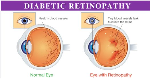
Diabetic Retinopathy (DR) is a common complication of diabetes that affects the eyes and can lead to vision loss. Detecting diabetic retinopathy typically involves analyzing images of the retina for signs of damage. Here's a general overview of how you can approach diabetic retinopathy detection using technology:
1. Image Acquisition
- Fundus Photography: High-resolution images of the retina are captured using specialized cameras known as fundus cameras or retinal cameras.
- Optical Coherence Tomography (OCT): Provides cross-sectional images of the retina to detect subtle changes.
2. Image Preprocessing
- Normalization: Adjust the brightness and contrast of the images to ensure uniformity.
- Noise Reduction: Apply filters to reduce noise and artifacts.
- Segmentation: Identify and isolate the region of interest (the retina) from the background.
3. Feature Extraction
- Detection of Retinal Features: Extract features such as blood vessels, microaneurysms, hemorrhages, and exudates.
- Texture Analysis: Analyze the texture of retinal images to identify abnormal patterns.
4. Classification
- Machine Learning Models: Train models using labeled datasets (images with known diagnoses) to classify images into categories such as no DR, mild DR, moderate DR, severe DR, or proliferative DR.
- Deep Learning: Convolutional Neural Networks (CNNs) are commonly used for automatic feature extraction and classification in retinal images.
5. Post-Processing
- Result Interpretation: Combine the results from classification models with clinical data to make diagnostic recommendations.
- Reporting: Generate reports with visualizations and recommendations for further action.
Additional information
Reviews
Add a Review
Your email address will not be published. Required fields are marked *


